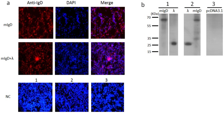
Figure 6. Immunofluorescence staining using the 13C2 mAb.
(a) Immunofluorescence staining of full-length mIgD heavy chain or λ light chain expressed on HEK293T cells with mAb 13C2(anti-IgD) plus goat anti-mouse IgG (red) and DAPI (blue). mIgD, cells transfected with full-length mIgD plasmid only; mIgD+λ, cells transfected with full-length mIgD heavy chain and λ light chain plasmids together. NC, negative control. 1, Cells transfected with an mIgD plasmid, stained by the secondary antibody goat anti-mouse IgG. 2, Cells transfected with mIgD and λ plasmids, stained by the goat anti-mouse IgG. 3, Cells transfected with the empty vector pcDNA3.1(+), stained by 13C2 plus goat anti-mouse IgG. Original magnification, ×20. (b) Immunoblot analysis of bovine mIgD heavy and light chains in cell lysates. mAb 13C2 was used to detect mIgD, and anti-FLAG antibody was used to detect the λ light chain. 1, Cell lysates from HEK293T cells transfected with mIgD heavy chain and λ light chain plasmids separately. 2, Cell membrane proteins extracted from HEK293T cells transfected with mIgD and λ plasmids together. 3, Negative control of membrane proteins from HEK293T cells transfected with empty vector pcDNA3.1(+).

