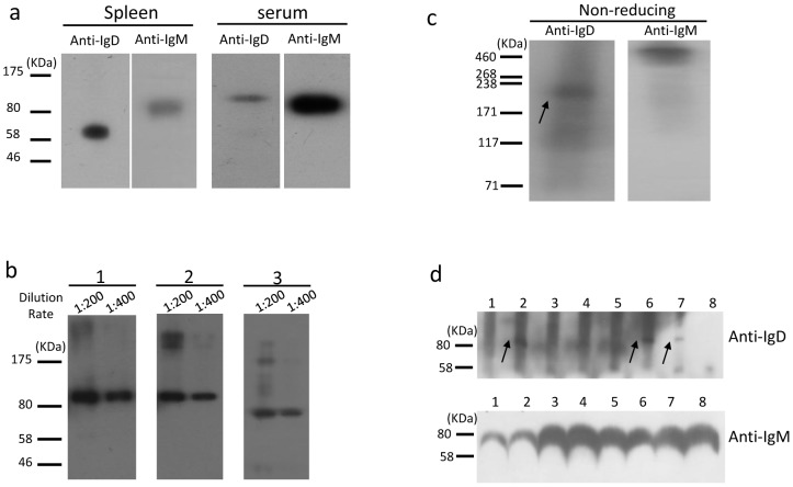
Figure 7. Immunoblot analysis of endogenous bovine IgD.
(a) Western blot of splenic membrane and serum proteins. (b) Western blot analysis of the IgD glycosylation in bovine serum. 1, Untreated serum proteins; 2, endo-α-N-acetylgalactosaminidase-treated serum proteins; 3, PNGase-treated serum proteins. The primary antibody 13C2 was diluted at 1∶200 or 1∶400. (c) Serum immunoblot analysis with mAb 13C2 and anti-bovine IgM polyclonal antibody under nonreducing conditions. The arrow indicates the monomer of IgD. (d) Immunoblot detection of serum IgD and IgM in differently aged cows. 1, 60 days old; 2, 180 days; 3, 1 year; 4, 2 years; 5, 3 years; 6, 4 years; 7, 5 years; 8, 6 years. The arrows indicate the IgD heavy chain.

