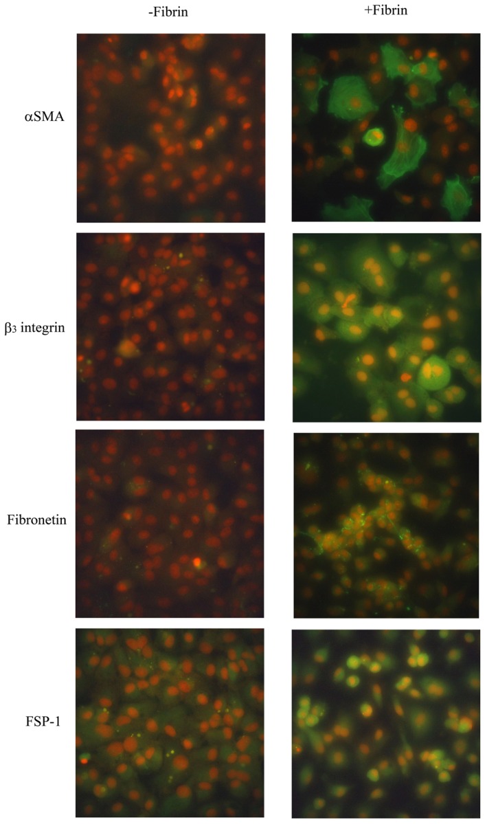
Figure 2. Changes in cell markers after application of fibrin to peritoneal mesothelial cells (PMCs).

Expression of α-smooth muscle actin (α-SMA), fibronectin, fibroblast specific protein-1 (FSP-1), and β3 integrin were detected by immunofluorescence staining with FITC-labeled secondary antibodies (green). Nuclei were counterstained with PI (red). PMCs overlaid with fibrin for 4 h (+Fibrin) expressed higher levels of α-SMA, fibronectin, FSP-1, and β3 integrin than untreated PMCs (-Fibrin). Original magnification ×400.
