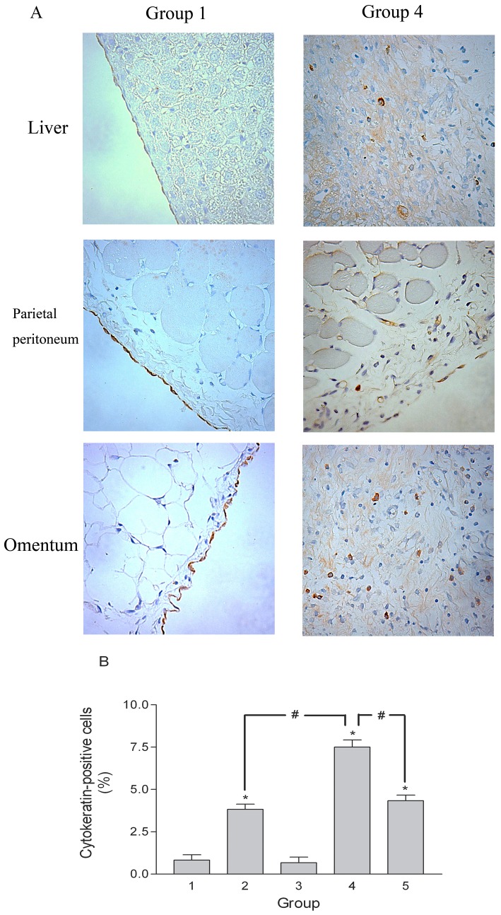
Figure 8. Immunohistochemical staining of cytokeratin.
(A) Cytokeratin-positive cells were present not only on the surface mesothelium, but also in the submesothelial fibrosis on the liver surface, parietal peritoneum, and omentum of Group 4 rats. Cytokeratin-positive cells were present only on the surface mesothelium of Group 1 rats. Original magnification ×400. (B) Percentage of cytokeratin-positive cells in the fibrotic tissue of the omentum. *p<0.05 vs. Group 1, #p<0.05.

