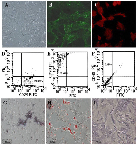
Figure 1.

Morphology and immunophenotypic characterization of MSCs. (A) The fibroblastic morphology of passage 3 MSCs (magnification = ×100); (B) MSCs stained with FITC-conjugated CD29 antibody (×200); (C) MSCs stained with PE-conjugated CD90 antibody (×200); (D), (E), and (F) MSCs analyzed by FACS for the positive expression of CD29 (D) and CD90 (E) and negative expression of CD45 (F); (G) Differentiated osteoblasts tested with alkaline phosphatase staining (×100); (H) Differentiated adipocytes characterized by oil red O staining (×100); (I) Differentiated chondrocytes verified by toluidine blue staining (×100).
