Figure 3.

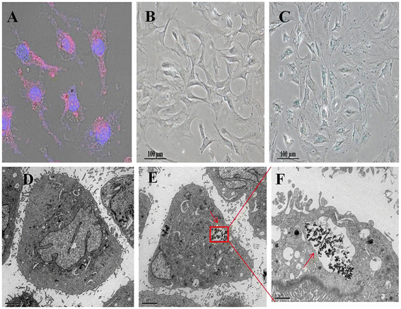
Evaluation of FMNP-labeled MSCs. (A) Cell nucleuses and FMNPs were visible in the MSCs by fluorescent microscope observation (×200); (B) FMNP-labeled MSCs were detected by using Prussian blue staining (×100); (C) Unlabeled MSCs were detected by using Prussian blue staining (×100); (D) The TEM image of unlabeled MSCs. (E) The TEM image of FMNP-labeled MSCs illustrates that amino-modified FMNPs were randomly distributed in the cytoplasm of MSCs (×6,000). (F) The magnification image of FMNPs distributed in the cytoplasm of MSCs (×12,000).
