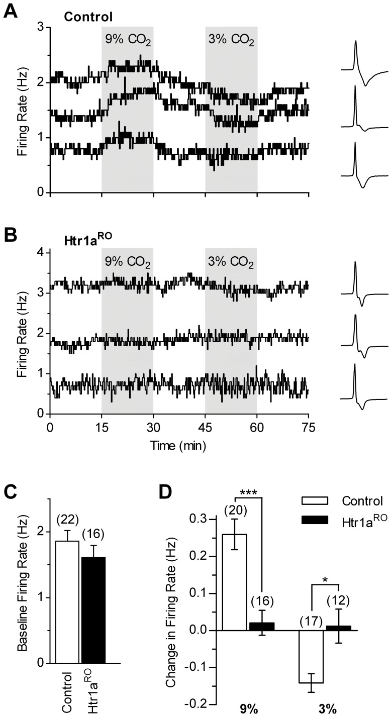
Figure 1. Intrinsic chemosensitive responses of serotonergic DRN neurons are greatly decreased in Htr1aRO mice.
A, B, Representative loose-seal cell-attached voltage-clamp recordings performed in the presence of synaptic blockers (see results) showing time-courses of serotonergic neuron firing in response to bath application of 9% and 3% CO2 in slices from control (A) and Htr1aRO (B) mice. Each panel reports three time-courses from different neurons. In B, one neuron with basal firing rate higher than the average of Htr1aRO group is shown to illustrate that the lack of responses to CO2 changes did not depend on basal firing rate of the recorded neuron (see results). Lines show firing rate calculated over 10 s bins. Traces illustrate recorded action currents for each experiment. C, Bar graph of baseline firing rate in the two groups. D, Summary bar graph comparing the effects of 9% and 3% CO2 in control and Htr1aRO mice. * p<0.05; *** p<0.001 (Mann-Whitney test). Number of recorded neurons is indicated in parentheses.

