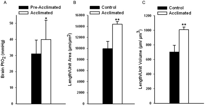
Figure 1. Partial pressure of oxygen in tissue (PtO2) measurements in brain parenchyma and capillary density data showing increased oxygen delivery after acclimation. A.
Brain (cortex) PtO2 measurements from the same animals both pre- and post-acclimation (n = 5, mean±S.D. paired t-test comparing pre- and post-acclimation, p<0.05). B. Capillary length per area of cortex before and after acclimation C. Capillary length per volume of cortex before and after acclimation. Measurements are from the dorsolateral cortex in the CA4-CA5 region and show the increased capillary density associated with acclimation to hypoxia. Control, non-acclimated subjects are compared with acclimated subjects (mean±S.D., *p<0.05, **p<0.005).

