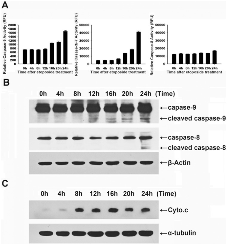
Figure 4. Etoposide induces apoptosis through caspase-9 and caspase-3 activation, mediated by mitochondrial cytochrome c release.
HeLa cells were treated with etoposide (50 µg/mL) for the indicated times. (A) Cell-free caspase-3, -8, and -9 activities were analyzed using specific substrates (Ac-DEVD-AFC, Ac-IETD-AFC, and Ac-LEHD-AFC, respectively). (B) The cells were analyzed by immunoblotting for caspase-8 and caspase-9. (C) Equal amounts of proteins from the cytosolic fraction were resolved by SDS-PAGE and analyzed by immunoblotting using antibodies against cytochrome c and α-tubulin.

