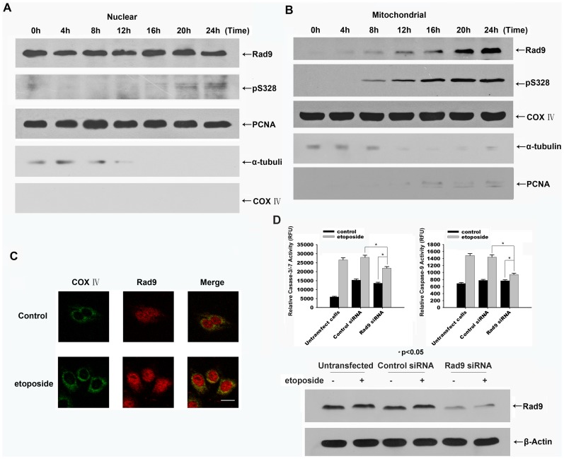
Figure 5. Serine 328-phosphorylated Rad9 is translocated from the nucleus to the mitochondria during etoposide-induced apoptosis in HeLa cells.
HeLa cells were treated with etoposide (50 µg/mL) for the indicated times. Equal amounts of protein from the nuclear fractions (A), and mitochondrial fractions (B) were resolved by SDS-PAGE and analyzed by immunoblotting using antibodies against Rad9, phospho328-Rad9, PCNA, α-tubulin, and COX IV. (C) The cells were treated with etoposide(50 µg/mL) for 20 h. The cells were fixed and stained with anti-Rad9 and anti-COX IV antibodies and analyzed by confocal microscopy using appropriate filters for the visualization of green, red, or combined fluorescence resulting from the presence of FITC and rhodamine molecules. Bar, 20 µm. (D) HeLa cells were transfected with negative control or Rad9 siRNA followed by the treatment with etoposide(50 µg/mL) for 20 h. Top: Cell extracts were assayed for caspase-3 and caspase-9 activities using the specific substrates Ac-IETD-AFC and Ac-DEVD-AFC (* p<0.05). Bottom: The contents of Rad9 and Actin in cell lysates were examined by immunoblotting.

