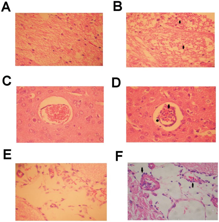
Figure 3. Pathological brain tissue alterations in mice infected with the JHA1 strain.
(A) Mock-treated mice with preserved brain parenchyma with typical cellular components. (B) Brain tissue of JHA1-infected mice showing a gliosis scar, in which dead neurons were replaced by astrocytes. The gliosis scar is indicated by the arrows. (C) Brain blood vessel (transverse view) in mock-treated mice. (D) Brain blood vessel in JHA1-infected mice filled with mononuclear cells (arrow). Some of the cells are observed outside the endothelial epithelium (asterisk) and are present in the brain parenchyma (arrowheads). (E) Brain blood vessel (longitudinal view) in mock-treated mice. (F) Brain blood vessel of JHA1-infected mice with infiltration of mononuclear cells (arrowheads). Infected and mock-infected mice were euthanized on day 8 p.i. Brain tissue samples were fixed with 1% formaldehyde, processed and stained with hematoxylin and eosin. Magnification: 400x. Images are representative of three independent experiments.

