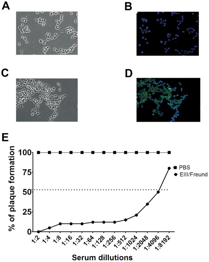
Figure 5. The JHA1 and NGC strains share immunological determinants within the major envelope protein.
LLC-MK2 cells were infected with the JHA1 strain and probed with a serum pool raised in mice immunized with EIII derived from the NGC strain or in non-immune animals. (A) Phase-contrast microscopy of the infected cells probed with serum from sham-treated mice. (B) Immunofluorescence microscopy of the infected cells probed with serum from sham-treated mice. The photograph was merged with the same field observed using phase-contrast microscopy. (C) Phase-contrast microscopy of infected cells probed with the anti-EIII serum pool. (D) Immunofluorescence microscopy of infected cells probed with the serum pool of mice immunized with the EIII protein derived from the NGC strain JHA1. The picture was merged with the same field observed in phase contrast. (E) Virus neutralization assay performed with the serum from JHA1-infected mice immunized with the EIII protein derived from the NGC strain. Aliquots containing 40 PFU were incubated with different dilutions of the anti-EIII serum pool (ranging from 1∶2 to 1∶8,192) for 30 min and subsequently transferred to wells with LLC-MK2 cells. One week later, the number of virus plaques was counted. Magnification: 400x. Images are representative of three independent experiments.

