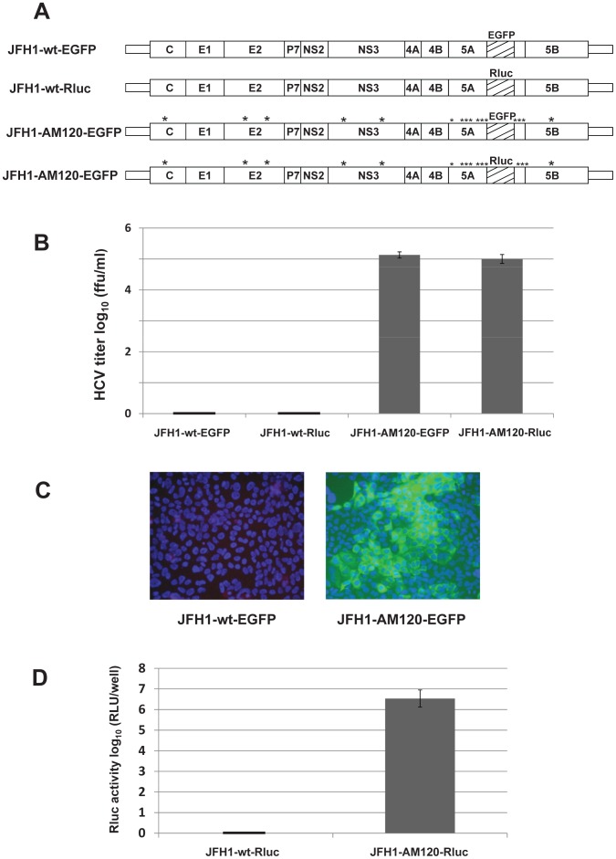
Figure 6. JFH1-AM120 HCV reporter viruses with gene inserts into NS5A; JFH1-AM120-Rluc and JFH1-AM120-EGFP.
(Panel A) Schematics of the HCV JFH1-wt and JFH1-AM120 based reporter constructs. EGFP and Renilla luciferace (Rluc) genes were inserted in frame into a unique RsrII site located in the amino acid 397 codon of JFH1-wt and JFH1-AM120. (Panel B) In vitro transcribed RNAs were electroporated into Huh-7.5 cells. Transfected cells were passaged for six days, infectivity titers of culture supernatants were measured and viral titers are expressed as focus-forming units per milliliter (f.f.u./ml) (triplicates that were performed twice). The data are presented as mean ± standard deviation (n = 6). (Panel C) JFH1-AM120-EGFP virus compared to JFH1-wt-EGFP. Huh-7.5 cells were inoculated with supernatants collected at six days post-transfection. Cells were fixed at 48 h post-infection and infected cells were identified by fluorescence immunostaining and microscopy as in Figure 3. Nuclear DNA was stained with DAPI (blue). (Panel D) Renilla luciferase activity was measured in Huh-7.5 cells following infection with day six supernatants from cells transfected with JFH1-wt-Rluc and JFH1-AM120-Rluc RNA. Assays were done in triplicate and in two separate experiments. The data are shown as mean ± standard deviation (n = 6).

