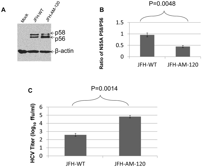
Figure 8. Phosphosphorylation of NS5A during HCV JFH1-WT and JFH1-AM-120 replication.
Huh7.5 cells were transfected with JFH1-wt or JFH1-AM-120 RNA as described in Methods. (Panel A) After three days of culture cells were lysed for Western blotting using anti-NS5A and anti-β-actin bodies. Western blots of proteins separated by 8% SDS-PAGE gel were done as described in Methods and the p56 and p58 isoforms of NS5A were identified. (Panel B) The quantyof p56 and p58 were determined using Image J software and the ratios of p58/p56 are shown. (Panel C). Virus titers in cell culture supernatant collected at the same times post infection (48 hours) were measured by determining focus-forming units with NS5A Immunofluorescence assays (Methods). Assays were done in duplicate and performed four different times. The data arepresented as mean ± standard deviation (n = 8). P-values were calculated using the student’s t-test.

