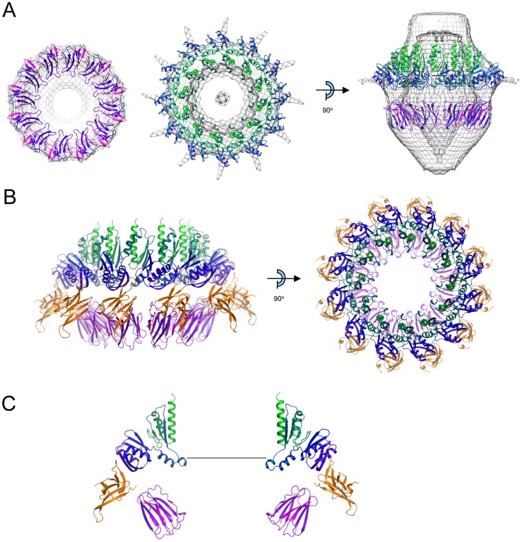
Figure 7. Model for the PilP-PilQ assembly.
A) Docking of B2 domain (left panel), N0/N1 domain (middle panel) and both (right panel) into the PilQ cryoelectron density map, contoured at 2.9σ. Some parts of the density map and oligomers have been removed for clarity. Colors are as used in Figure 2A (B2 domain) and Figure 3B (N0/N1 domains). B) Reconstruction of PilQ N0/N1/B2 domain structures (colored as in A) with PilP C-domain (orange) bound. Left panel: side view with 6 oligomers; right panel: top view with 12 oligomers. C) Detail of two oligomers on opposing sides of the PilQ chamber. The scale bar is 60 Å and corresponds to the approximate dimensions of an assembled type IV pilus fiber.

