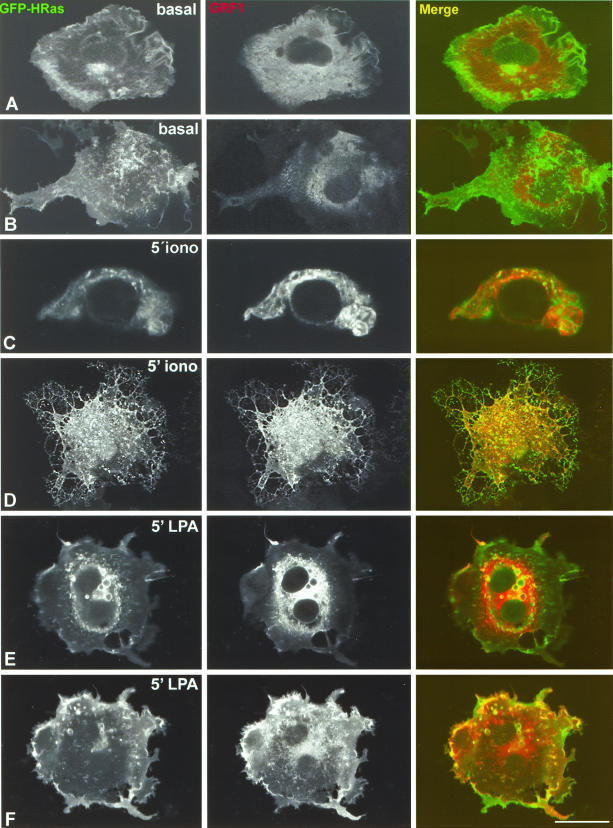
FIG. 4.
Colocalization of RasGRF1 and H-Ras under stimulation. COS-7 cells were cotransfected with suboptimal concentrations of RasGRF1 (0.1 μg) and with GFP-H-Ras (0.25 μg) and analyzed using confocal microscopy. (A and B) Basal conditions. An equatorial confocal section at the level of the cell nucleus (A) and a tangential section illustrating the PM (B) are shown. (C and D) Treatment with 1 μM ionomycin for 5 min. A confocal section at the cell nucleus level (C) and a confocal section sweeping through the cell surface (D) are shown. (E and F) Treatment with 10 μM LPA for 5 min. A confocal section at the cell nucleus level (E) and a confocal section illustrating the cell surface (F) are shown. Bars, 10 μm (5 μm for panel C).

