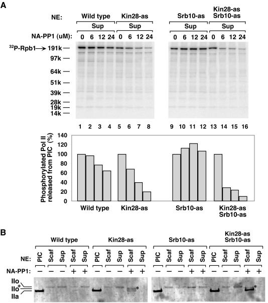
FIG. 6.
Inhibition of Kin28 and Srb10 kinases inhibits phosphorylation of the Pol II CTD. (A) Phosphorylated Pol II released into the supernatant (Sup) during Scaffold complex formation. Phosphorimager analysis was performed on the membrane containing the supernatants (Fig. 5), and the quantitation of the phosphorylated Pol II CTD is shown below. The phosphorylation signal of each extract without inhibitor (lanes 1, 5, 9 and 13) was normalized to 100% (lower panel). (B) Both Kin28 and Srb10 contribute to hyperphosphorylation of the CTD during Scaffold complex formation. PIC and Scaffold complexes formed from the indicated extracts were fractionated on a 3 to 8% Tris-acetate gel and analyzed by Western blotting using antibody YN-18, which detects Rpb1 independent of the phosphorylation state. IIo* represents a partially phosphorylated Pol II form.

