
Figure 6.
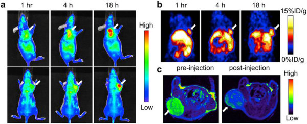
In vivo NIRF (a) and PET (b) images of mouse injected with iron oxide nanoparticles. Images were acquired 1, 4, and 18 h after injection. (c) MRI images acquired before and 18 h after injection. Reproduced with permission from [73].
Official websites use .gov
A
.gov website belongs to an official
government organization in the United States.
Secure .gov websites use HTTPS
A lock (
) or https:// means you've safely
connected to the .gov website. Share sensitive
information only on official, secure websites.
In vivo NIRF (a) and PET (b) images of mouse injected with iron oxide nanoparticles. Images were acquired 1, 4, and 18 h after injection. (c) MRI images acquired before and 18 h after injection. Reproduced with permission from [73].