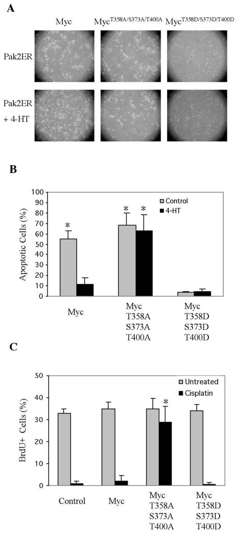
FIG. 9.
Activation of Pak2 reduces apoptosis induced by Myc. (A) NIH 3T3 cells expressing Pak2ER were infected with a retroviral vector expressing various alleles of MYC. At 24 h after infection, cells were treated for 12 h with 4-HT. They were then kept in serum-free medium with 4-HT for 4 days. Induction of Pak2 activity reduced apoptosis induced by Myc. Photomicroscopy was performed at a magnification of ×40. (B) Quantification of apoptotic cells in panel A. Apoptotic cells were labeled by 7-aminoactinomycin D staining and analyzed by flow cytometry. The histograms represent the mean of three experiments, with error bars indicating standard deviation (*, P < 0.01). (C) A Myc mutant that cannot be phosphorylated by Pak2 drives cell cycle progression under stress. NIH 3T3 cells expressing various alleles of Myc were treated for 24 h with cisplatin, labeled with BrdU for 30 min, and analyzed as described in Materials and Methods. The histograms represent averages from three experiments, with error bars indicating standard deviation (*, P < 0.01).

