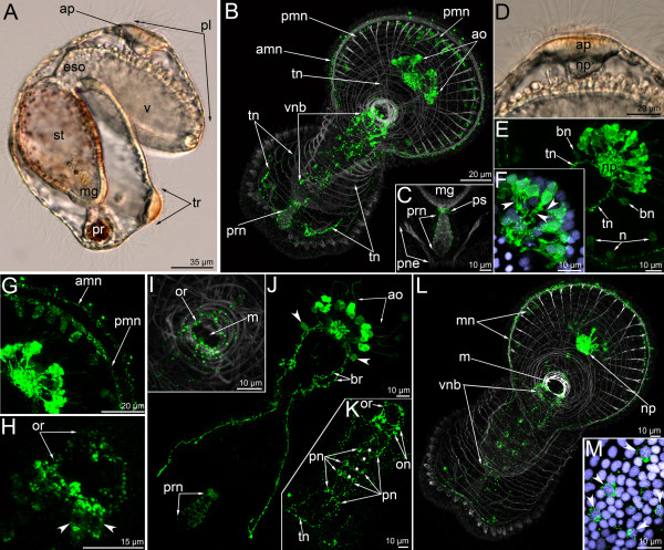
Figure 6.
The organization of the serotonin-like immunoreactive nervous system in a 6-day-old larva. Micrographs of live animals (A) and Z-projections of larvae after mono- and double staining with antibodies against serotonin (green), as well as with phalloidin (grey), and Hoechst (violet). Apical is to the upper right on all micrographs except A, C, and D, where apical is to the top. (A) Lateral view with ventral to the right. Larva with apical plate (ap), preoral lobe (pl), vestibulum (v), esophagus (eso), stomach (st), midgut (mg), proctodaeum (pr), and tentacular ridge (tr). (B) Overview of the musculature and serotonin-like immunoreactive nervous system showing apical organ (ao), anterior marginal neurite bundle (amn), posterior marginal neurite bundle (pmn), tentacular neurite bundle (tn), and ventral neurite bundles (vnb). Ventral view; preoral lobe bends backward. (C) Posterior body part. The micrograph comprises selected optical sections from the mid body region of the specimen and shows the nerve net around the proctodaeum (prn) and the pyloric sphincter (ps), midgut (mg), and protonephridia. (D) Lateral view of the apical plate (ap) and large neuropile (np) underneath. The anterior pole of the preoral lobe is to the right. (E) Top view of the apical organ with dorsal neuropil (np), dorso-lateral branches of the tentacular neurite bundle (tn), and bipolar perikarya (bn). Some portions of the marginal neurites with perikarya (n) are visible. (F) Details of the apical organ, anastomosing neurites are indicated by arrowheads. (G) Anterior portion of the edge of the preoral lobe with anterior (amn) and posterior (pmn) marginal neurite bundles with a group of perikarya associated with the latter. (H) Oral nerve ring with ventro-lateral, weakly stained perikarya (arrowheads); top view. (I) Innervation (oral nerve ring – or) and musculature of the mouth (m); ventral view. (J) Dorsal portion of the tentacular neurite bundle (tn) with two branches (br) of the right stem. apical organ (ao), bipolar perikarya (arrowheads), and nerve net of the proctodaeum (prn). (K) Oral field with the ventral neurite bundles, which pass from the oral ring (or) with oral perikarya (on) to the tentacular neurite bundle (tn), containing several paired perikarya (pn) and commissures (asterisks). The micrograph is composed of the most ventral optical sections only. (L) The image comprises only the four most ventral optical sections of the serotonin labeling and all sections of the muscle staining. Ventral view of a larva showing the marginal neurites (mn) of the preoral lobe, the neuropil (np) of the apical organ, the mouth (m), and the ventral neurite bundles (vnb). (M) Some paired perikarya of the ventral neurite bundles with nuclei (arrowheads).

