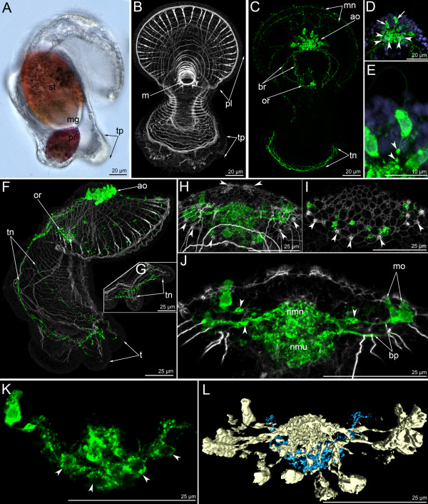
Figure 7.
The organization of the serotonin-like immunoreactive nervous system in a 13-day-old larva. Micrographs of a live animal (A), Z-projections (B-K) of larvae after mono- and double staining with antibodies against 5-HT (serotonin) (green), as well as staining with phalloidin (grey), and Hoechst (violet), and 3-D reconstruction (L). Apical is to the top in all aspects. (A) Lateral view of live larva showing stomach (st), midgut (mg), proctodaeum (pr), and primordia of tentacles (tp). (B) Overview of the muscle system; ventral view; preoral lobe bends backward. General view showing preoral lobe (pl), mouth (m), and primordia of tentacles (tp). (C) The same larva; overview of the serotonin-likeimmunoreactive system; ventral view showing weakly stained marginal nerves, apical organ (ao), branches (br) of the dorsal portion of the tentacular neurite bundle, oral ring (or), and ventral portion of the tentacular neurite bundle (tn). (D-E) Details of the apical organ; anterior view. (D) General view of the apical organ, which is composed of monopolar (arrows) and bipolar or multipolar (arrowheads) perikarya. (E) Monopolar perikaryon with varicosities (arrowheads) on the basal process. (F) Overview of the musculature and the serotonin-like immunoreactive nervous system showing apical organ (ao), oral ring (or), tentacular neurite bundle (tn), and tentacles (t). Lateral view; ventral is to the right. (G) Organization of the ventral portion of the tentacular neurite bundle (tn) which forms a loop in each tentacle. (H) Ventral view of the apical plate with apical organ and rows of sensory cells (arrowheads). (I) The micrograph comprises the three ventral-most optical sections of the confocal stack, i.e., the region of the mutual position of monopolar perikarya and sensory cells (arrowheads). (J) Frontal view of the center of the apical organ showing monopolar perikarya (mo), their basal processes (bp) with varicosities (arrowheads) and neuropil (nmn), and bipolar or multipolar perikarya and neuropil (nmu). (K) Organization of bipolar or multipolar perikarya (arrowheads); ventral view. (L) Three-dimensional reconstruction of the apical organ. Light blue indicates multipolar or bipolar perikarya, which form a horseshoe-shaped row under the neuropil of the monopolar perikarya (golden); ventral view.

