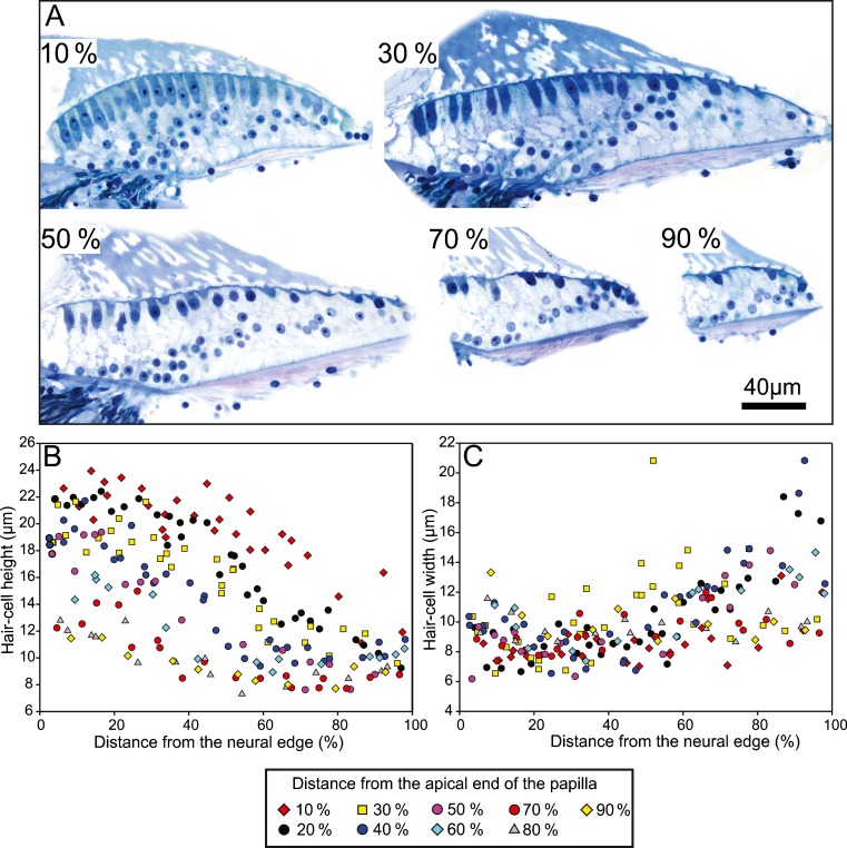
Fig. 3.
Hair cell morphology in the kiwi basilar papilla. A High magnification images of the basilar papilla at 10 %, 30 %, 50 %, 70 %, and 90 % positions from the apical end of the papilla. Height (B) and width (C) of hair cells in the kiwi basilar papilla as a function of the normalized distance from the neural edge of the papilla. Symbols represent the position of the hair cells along the length of the basilar papilla as indicated in the legend.

