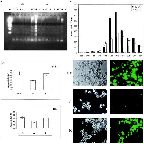
FIG. 7.
Decreased sensitivity of Gab1−/− cells to UV-induced apoptosis. Fibroblasts were stimulated by UV-B irradiation (400 J/m2) and incubated at 37°C for the indicated times. (A) Fragmented DNA was extracted and analyzed by electrophoresis on a 1.5% agarose gel. M, molecular marker; C, control. (B) Caspase 3 activity was determined as the rate of product formation obtained from the linear portion of the reaction curves within the first 10% of substrate depletion. Values represent the mean and standard deviation of three independent experiments. (C) Wild-type, Gab1−/−, and Gab1-transfected Gab1−/− cells (R) were irradiated with UV-B (400 J/m2). After incubation at 37°C for 18 or 48 h, cell apoptosis was evaluated using the YOPRO-1 staining kit. Apoptotic cells were counted under fluorescent optics, and the total cell number in the fields was counted under phase optics. The percentage of apoptotic cells was calculated, and the data were averaged from three samples (means and standard deviations are shown). Shown on the right are representative microscopic fields of cells at 48 h under phase optics (left) and fluorescent optics (right). +/+, wild-type cells; −/−, Gab1−/− cells.

