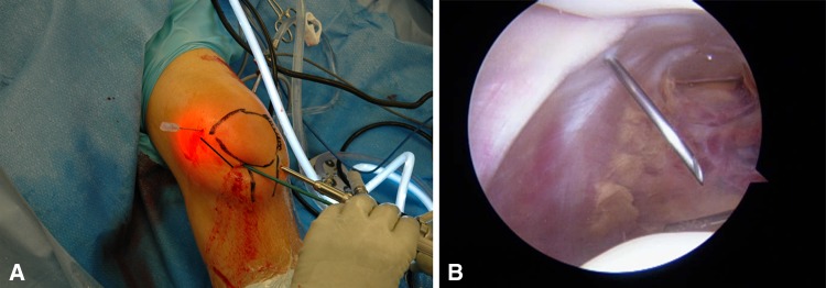
Fig. 2A–B.
The surgeon should correlate the (A) external and (B) internal anatomy to ensure that excessive release is not done proximally. In Illustration A, a line is drawn depicting the course of a lateral release. Illustration B shows the muscle fibers of the vastus lateralis proximal to the spinal needle.

