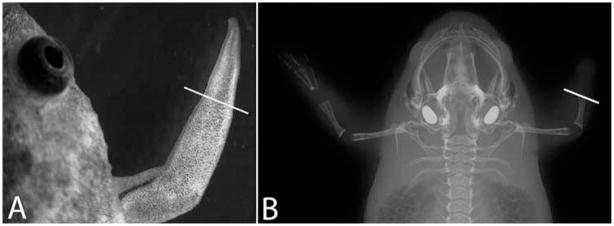
Fig. 1.
X-ray images of Xenopus frogs. A: A bright field image of a regenerated frog spike. B: X-ray image of a frog captured by the In Vivo MS FX PRO imaging system. Limb amputation was performed on the right forelimb only and a spike was formed on the right amputation stump. The white lines indicate the amputation planes.

