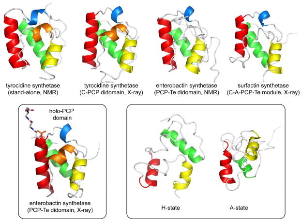
Fig. 3.
Representative structures of PCP domains from NRPS assembly line constructs. (a) Structures of apo-PCP domains, PDB codes: 2GDW, 2JGP, 2ROQ, 2VSQ (left to right). (b) Structure of a holo-PCP domain showing site of phosphopantethiene attachment, PDB code, 3TEJ. (c) Alternate conformations of a tyrocidine PCP domain observed in the context of binding partners, PDB codes, 2GDX, 2GDY. The N- and C-termini of the domains are indicated.

