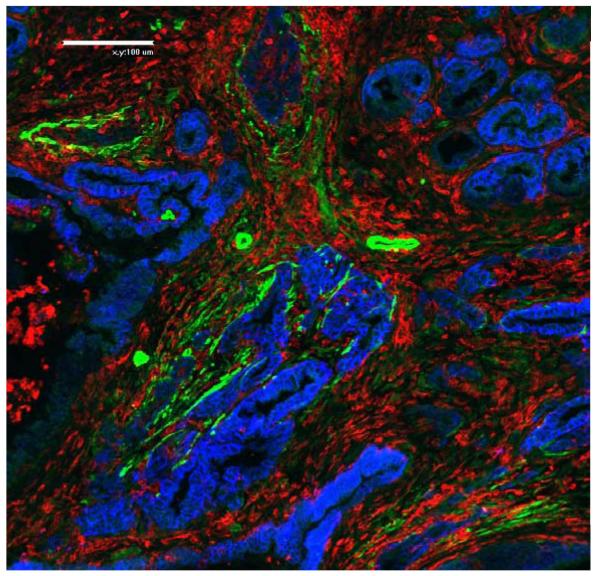
Fig.3.
Prominent immune cell infiltration exists in mouse PDA. This immunofluorescence staining illustrates the abundance of immune cells marked by CD45 expression (red) between neoplastic glandular structures (stained for EpCam in blue) and α-SMA positive stromal fibroblasts and perivascular cells that likely represent pericytes (green) (20x magnification).

