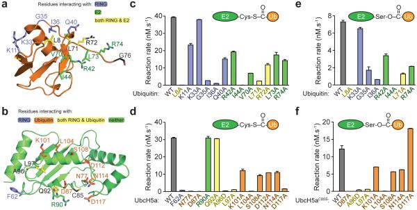
Figure 3. Mutational analysis of the RNF4 RING–UbcH5a~Ub complex.
a, Side chains of altered residues in ubiquitin contacting RNF4 (blue), UbcH5a (green), or both RNF4 and UbcH5a (yellow). b, Side chains of altered residues in UbcH5a contacting RNF4 (blue), ubiquitin (orange), both RNF4 and ubiquitin (yellow), or neither (green). c, Reaction rates were determined (mean ± s.d. of duplicates) for single-turnover, RNF4-dependent substrate ubiquitylation assays with mutant forms of ubiquitin. Wild-type ubiquitin is in grey and mutants are colour coded as in a. d, Assays with UbcH5a mutants quantified as in c and colour coded as in b. e, RNF4-mediated hydrolysis of UbcH5aC85S~Ub oxyesters with mutations in ubiquitin. Rates are mean ± s.d. of duplicates. f, As in e, with mutations in UbcH5a.

