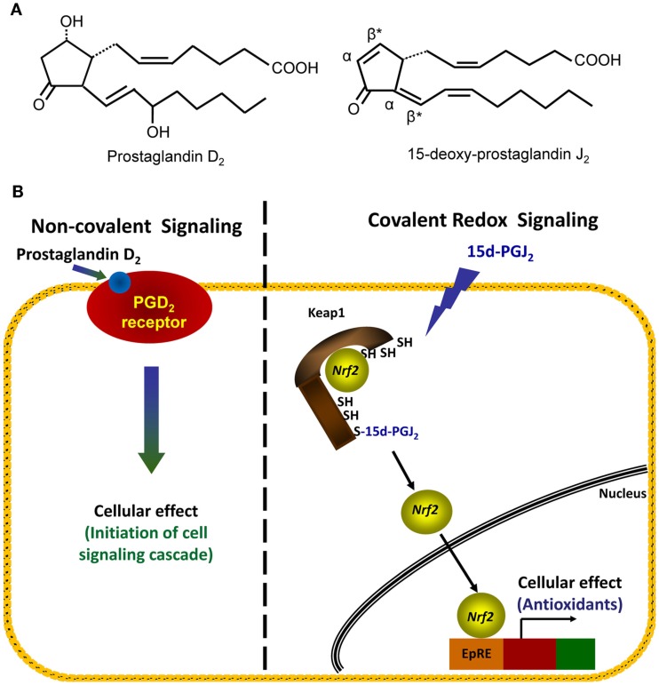
Figure 3.
Comparison of non-covalent and covalent signaling molecules. (A) The structures of two prostaglandins, PGD2 (left structure) and 15d-PGJ2 (right structure), are shown. Electrophilic carbons which arise from presence of α, β-unsaturated carbonyl functional groups are denoted by asterisks, and α- and β-carbons are indicated. (B) Left hand panel: in the non-covalent or classical receptor-ligand model (left hand panel), the ligand PGD2 is recognized by a specific receptor on the cell membrane. The binding event causes the activation of a signaling cascade which ultimately changes cellular function. Right hand panel: in the covalent signaling model, a reactive species 15d-PGJ2 directly forms a covalent adduct with select proteins, in this case, the cytosolic repressor protein Keap1. This binding event causes changes in endogenous antioxidant protein transcription via the transcription factor Nrf2.

