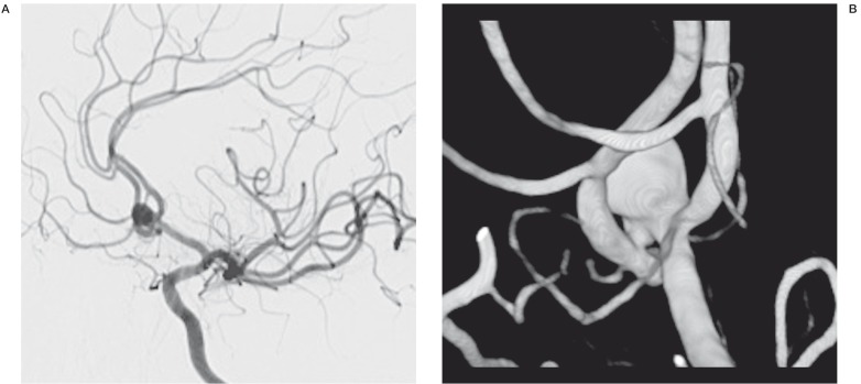
Figure 1.
A) The left ICA angiogram, oblique view, shows the AcomA aneurysm with a wide neck involving both A2 segments. B) Three-dimensional image of the AcomA aneurysm, showing the left A1 segment and the left and right pericallosal arteries. The right pericallosal artery is clearly emerging from the neck of the aneurysm.

