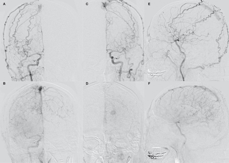
Figure 2.
A-D) Anterior-posterior view of a common carotid artery angiogram (A, right, arterial phase; B, right, late phase; C; left, arterial phase; D, left, late phase) demonstrating dural arteriovenous fistulas in the superior sagittal sinus. The feeders include the bilateral middle meningeal arteries, superficial temporal arteries, and occipital arteries. Severe venous congestion and a varix are observed; E,F) Lateral view of a right common carotid artery angiogram (E, arterial phase; F, late phase) demonstrating dural arteriovenous fistulas in the superior sagittal sinus. The posterior third of the sinus is occluded.

