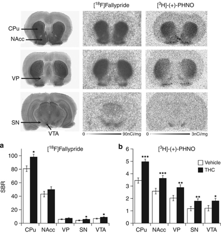
Figure 2.
Brain regional D2/3R availability (SBR) as measured with ex vivo autoradiography binding of [18F]Fallypride (a) and [3H]-(+)-PHNO (b) at 24 h following cessation of a 3-weeks treatment with either vehicle or THC. Data are shown as mean±SEM (n=8–10) in the caudate/putamen (CPu), nucleus accumbens (NAcc), ventral pallidum (VP), substantia nigra (SN), and ventral tegmental area (VTA). Significantly different from the vehicle-treated group at *p<0.05, **p<0.01, and ***p<0.001 using a mixed model repeated-measures ANOVA with a post hoc t-test. The upper panel shows representative photographs of acetylcholinesterase (Ach) staining and autoradiograms of [18F]Fallypride and [3H]-(+)-PHNO bindings obtained in saline-treated rats in comparable coronal sections taken at 1.6 mm (top row), −0.26 mm (middle row), and −5.60 mm (bottom row) relative to bregma of a rat brain atlas (Paxinos and Watson, 1998).

