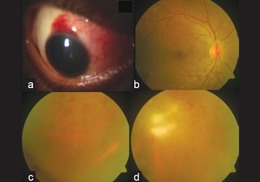
Figure 1.

Clinical signs at presentation. (a) Area of occult scleral injury marked by localized congestion and chemosis; (b) fundus photograph showing macular internal limiting membrane striae; (c) white-centered retinal hemorrhages; (d) midperipheral and peripheral retinal vasculitis
