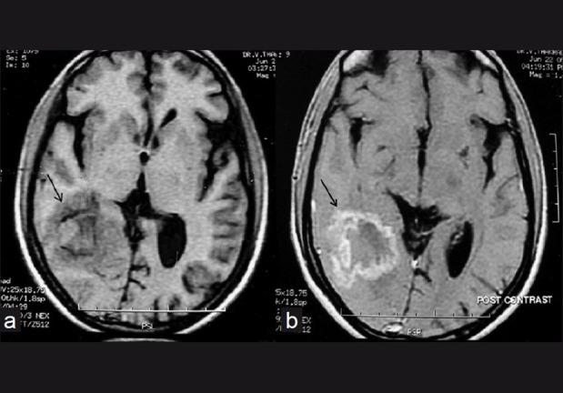
Figure 3.

Pre-treatment Axial T1W image (TR/TE-340/11) shows the lesion in the right temporo-occipital region (arrow) with a periphery which is isointense to grey matter, an incomplete hypointense rim and a mixed iso and hyperintense centre (a). Axial contrast-enhanced T1W image shows irregular ring enhancement of the lesion (arrow) (b)
