Figure 4.

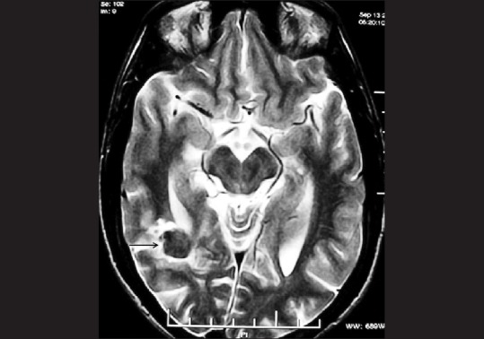
Follow-up axial T2W MRI after one year reveals a significant decrease in the size of the lesion (arrow) which now has a hypointense centre

Follow-up axial T2W MRI after one year reveals a significant decrease in the size of the lesion (arrow) which now has a hypointense centre