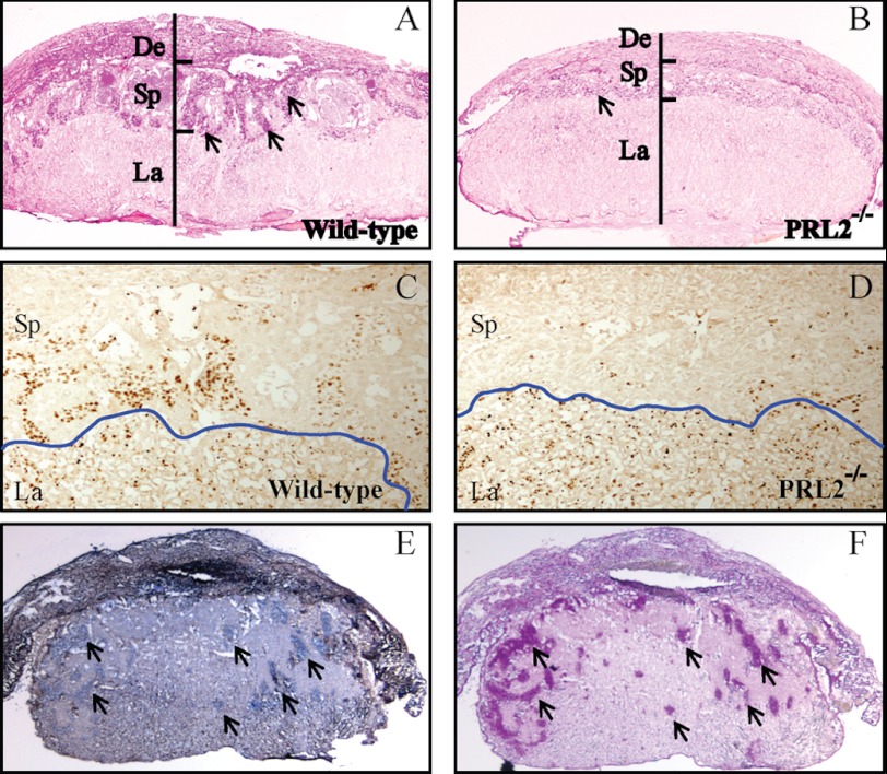
FIGURE 4.
PRL2 mutant placenta has significantly lower amount of spongiotrophoblast and glycogen cells due to reduced proliferation. A–D, histological examination of wild-type and PRL2−/− placentas at E12.5. A and B, PAS glycogen staining. Arrows designate islands of glycogen cells. C and D, immunohistochemistry using antibody against Ki67 to monitor proliferating cells. The blue line shows the boundary between labyrinth and spongiotrophoblast layers. De, deciduas; Sp, spongiotrophoblast; La, labyrinth. E, in situ hybridization using PRL2 probe showing PRL2 mRNA expression on frozen section of E12.5 wild-type placenta. Arrows show PRL2-positive cells in the decidual layer. F, PAS glycogen staining on the section adjacent to E. Arrows show glycogen-containing cells. Note the overlap of PRL2-positive cells and glycogen cells.

