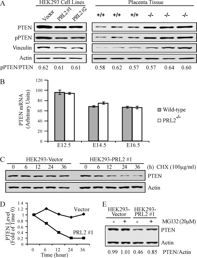
FIGURE 7.

PRL2 destabilized PTEN protein by reducing vinculin expression. A, lysates from E12.5 PRL2−/− or wild-type placentas as well as HEK293-PRL2 stable cell lines (PRL2 #1 and #2) and their vector controls were analyzed by Western blot for PTEN, phospho-PTEN (pPTEN), and vinculin expression. B, PTEN mRNA levels in PRL2−/− or wild-type placentas at E12.5, E14.5, and E16.5 were determined by quantitative RT-PCR (n = 5 for each genotype at each stage). Data represent mean ± S.E. C, HEK293-PRL2 stable cell line and its vector control were treated with cycloheximide (CHX) at 100 μg/ml for the indicated times, and cell lysates were analyzed for PTEN and actin protein levels by Western blot. Data represent three independent experiments. D, quantification of the results in C. PTEN and actin signals were measured using ImageJ. The ratio of PTEN/actin was determined for each sample and plotted as -folds of time 0 for each cell line. E, HEK293-PRL2 cell line and its vector control were treated with 20 μm MG132 for 6 h. Cells were lysed, and PTEN and actin protein levels were analyzed by Western blot. PTEN and actin signals were measured using ImageJ, and the ratio of PTEN/actin was determined. Data are representative of three independent experiments.
