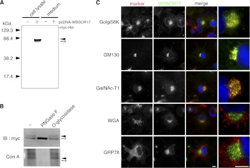
FIGURE 2.
Characterization of recombinant WBSCR17 protein in COS7 cells. A, the culture medium and cell lysates were collected and analyzed by Western blotting with an anti-Myc antibody. Only in cell lysate of the WBSCR17 transfectant, two bands of about 80 and 71 kDa were observed. Black and white arrowheads indicate the upper and the lower bands, respectively. B, treatment of N-glycosidase PNGase F, but not O-glycosidase, led to a decrease in the 80-kDa form and increase in the 71-kDa form (top). The upper band positive for ConA staining was lost upon digestion with PNGase F (bottom). Black and white arrowheads indicate the upper and lower bands, respectively. C, subcellular localization of recombinant WBSCR17 in COS7 cells. The recombinant WBSCR17 was detected with anti-Myc antibody, and its subcellular localization was compared with that of several organelle markers: Golgi58K, Golgi; GM130, cis-Golgi; GalNAc-T1, cis-Golgi and medial Golgi; WGA, trans-Golgi; and Grp78, ER. The rightmost panels are magnified images of the merged panels. WBSCR17 was predominantly localized in the Golgi apparatus. Scale bars, 10 μm. IB, immunoblot.

