FIGURE 4.

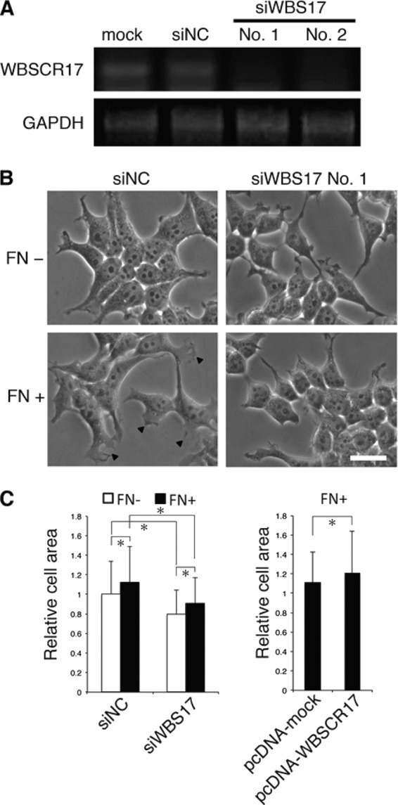
Suppression of WBSCR17 in HEK293T cells inhibits lamellipodium formation. A, validation of siRNA suppression by RT-PCR analysis. WBSCR17 mRNA levels were evaluated by RT-PCR using primers for WBSCR17. GAPDH mRNA was used as a control for equal gel loading. Two siRNAs against WBSCR17 (No. 1 and No. 2), but not control siRNA (siNC), suppressed the WBSCR17 mRNA expression. B, change of cell morphology by knockdown of WBSCR17. A dish coated with FN induced cell spreading and lamellipodium formation (bottom left, lamellipodia are indicated by arrowheads). HEK293T cells transfected with WBSCR17 siRNA remained round-shaped and did not form lamellipodia on the FN-coated dish. Scale bar, 20 μm. C, measurement of cell surface area revealed the involvement of WBSCR17 in cell spreading. Cell surface area was quantified using ImageJ software. Suppression of WBSCR17 led to decreased cell area, whereas WBSCR17 overexpression gave rise to an increased area. Data shown represent means ± S.D. (error bars) of three independent experiments. *, p < 0.05.
