FIGURE 5.
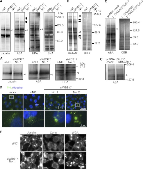
Alterations of O-glycan profiles and intracellular accumulation of glycoconjugates in the WBS17KD cells. A and A′, profiles of glycoproteins in the cell lysates from control and WBS17KD cells were examined with Jacalin, ABA, HPA, and SNA lectins. Black and white arrowheads indicate down-regulated and up-regulated glycoproteins, respectively. The magnified images of bands of ∼100 kDa that were up-regulated in the knockdown cells (A) are shown in A′. B, metabolic labeling of glycoproteins with GalNAz, an azide-modified GalNAc. Cell lysates were prepared from the cells that were labeled with GalNAz. GalNAz-labeled glycoproteins in cell lysate were chemically conjugated with tetramethylrhodamine, subjected to SDS-PAGE, and detected by fluorescence observation. Total proteins were stained by Coomassie Brilliant Blue (CBB). There were changes in O-glycan profiles in the WBS17KD cells. Black and white arrowheads indicate down-regulated and up-regulated O-glycans, respectively. C and C′, lectin blot analysis with ABA detected enhanced expressions of ∼150-kDa O-glycoproteins (indicated by an asterisk) in the cells with WBSCR17 overexpressed. C′, a magnified image of C. D and E, lectin staining of HEK293T cells with a PHL lectin (D) and Jacalin, ConA, and WGA lectins (E). Intracellular accumulations of glycoconjugates that were positive for all of the lectins used were detected in WBSCR17 siRNA transfectants. The bottom panels in both D and E are magnified images of the boxed areas in the top panels.

