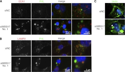
FIGURE 6.
Accumulation of glycoconjugates in the lysosomes in the WBS17KD cells. Two days after the transfection of siRNAs, HEK293T cells were stained with fluorescence-labeled PHL lectin and antibodies against organelle markers: EEA1 for early endosomes (A) and LAMP2 for lysosomes (B). The glycoconjugates positive for PHL accumulated in the vesicles that are labeled with LAMP2 but not with EEA1. The vesicles that were positive for either EEA1 or LAMP2 were enlarged in the WBS17KD cells. The rightmost panels are magnified images of the merged panels. C, cell surface proteins of HEK293T cells were biotinylated, the cells were cultured for 4 h, and then the biotinylated proteins were reacted with FITC-streptavidin and analyzed. The analysis revealed increased internalization of plasma membrane proteins to large vesicles (indicated by an arrowhead).

