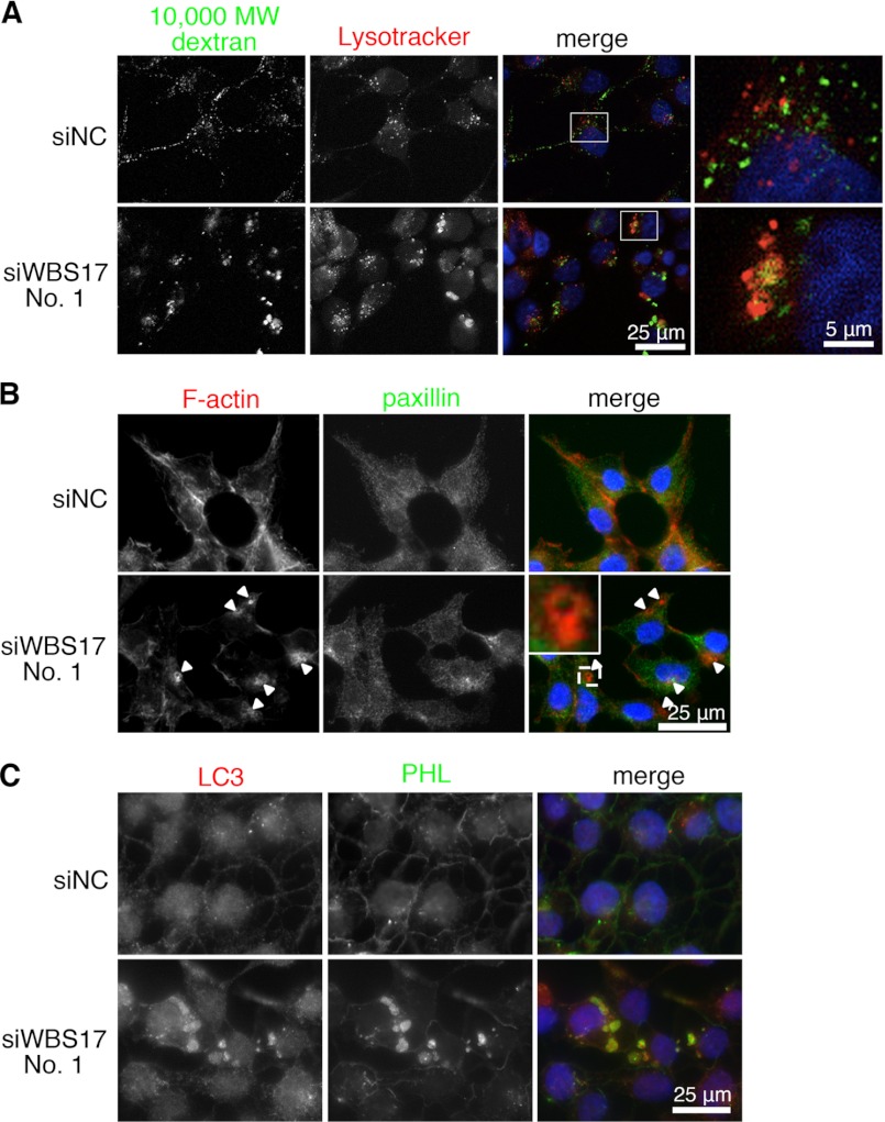
FIGURE 8.
WBSCR17 regulates macropinosome formation. A, co-labeling of the cells with Mr 10,000 dextran and Lysotracker, a lysosome marker. In the control cells, most of the vesicles positive for dextran were small and not stained with Lysotracker. The WBS17KD cells, on the other hand, had large vesicles co-labeled with dextran and Lysotracker. The rightmost panels are magnified images of the boxed areas in the merged panels. B, staining of HEK293T cells with phalloidin, which binds to F-actin, and with anti-paxillin antibody. The arrowheads indicate actin-rich round-shaped rufflings in the WBS17KD cells. The top left corner of the merged image of the knockdown cells is a magnified image of the boxed area. C, staining of HEK293T cells with antibody to LC3 and a PHL lectin. In the WBS17KD cells, LC3 was associated with the large vesicles positive for PHL staining.

