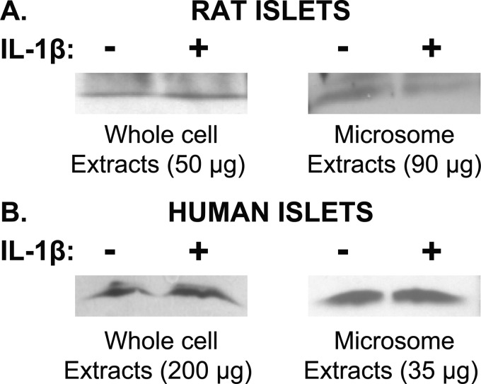
FIGURE 5.
Western analyses for mPGES-1 using microsomal versus whole cell extracts. Top panel, rat islets are shown. Whole cell extracts and microsomal extracts using greater amounts of protein than were used in Fig. 3 failed to reveal stimulation of mPGES-1 levels by IL-1β. Bottom panel, human islets are shown. mPGES-1 was detectable in whole cell extracts using greater amounts of protein than were used in Fig. 3 and revealed mPGES-1, but these levels were not increased when islets were treated with IL-1β.

