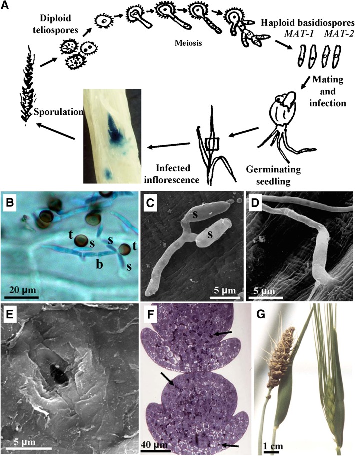
Figure 1.
Infection Process of U. hordei on Barley.
(A) Schematic representation of the U. hordei life cycle. Dispersed teliospores become lodged under seed hulls and germinate together with the seed. Mating precedes infection and occurs on the young coleoptile. The photographic insert depicts a light microscopy picture of an immature inflorescence at 47 d, colonized after infection by a compatible, mated mixture of U. hordei strain Uh359 stably transformed with a β-glucuronidase–expressing construct and strain Uh362. Fungal hyphae were colored blue following treatment of the inflorescence with glucuronide (Hu et al., 2002).
(B) Light microscopy picture of germinated teliospores (t) stained with cotton blue on the surface of a barley coleoptile 17 h after inoculation having produced a basidium (b), from which haploid basidiospores (s) are emerging. Microscopy procedure as described by Gaudet et al. (2010).
(C) Scanning electron micrograph of two mated basidiospores (s) of opposite mating type fused through conjugation hyphae to produce the dikaryotic infection filament on a barley coleoptile.
(D) Scanning electron micrograph of a dikaryotic infection hypha entering the barley coleoptile wall by direct penetration.
(E) Scanning electron micrograph showing a penetration site, from which the hypha was dislodged during sample preparation. Scanning electron microscopy in (C) to (E) as described by Hu et al. (2002).
(F) Light microscopy picture of an immature inflorescence showing extensive, early teliospore formation (arrows).
(G) Emerged barley inflorescence where all kernels have been replaced by black teliospores (left) next to a healthy head (right).

