Figure 6.
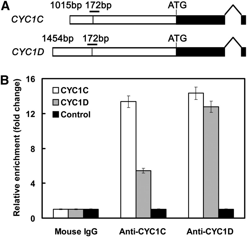
Enrichment of CYC1C and CYC1D Promoters by Anti-CYC1C and Anti-CYC1D Antibodies in ChIP Assays.
(A) The structure of P. heterotricha CYC1 genes. The white boxes represent sequences upstream of the start codon, with vertical lines indicating the position of the CYC binding sites. The horizontal bars above the binding sites indicate the fragments amplified in ChIP-qPCR experiments. The black boxes represent the coding regions.
(B) Enrichment of either the CYC1C or CYC1D promoter by both anti-CYC1C and anti-CYC1D antibodies. The coding sequence of CYC1C located at ∼1.5 kb downstream of the CYC binding site is amplified as a negative control. The data (mean ± sd) are determined from at least three fully independent experiments.

