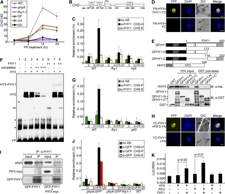
Figure 4.
FHY1 and phyA Are Recruited to the CHS Promoter through HY5 and PIF3 in FR.
(A) FHY1 phosphorylation inhibits FR-induced CHS expression. Four-day etiolated seedlings of indicated genotypes were exposed to FR (6 µmol/m2/s) for the indicated time. The values of CHS transcript were normalized to 18S rRNA in the quantitative RT-PCR. WT, wild type.
(B) A diagram of the CHS gene and promoter. CHS-H and CHS-P are HY5- and PIF3 binding regions containing G-boxes (indicated by the triangles). CHS-E is a region on an CHS exon.
(C) FHY1 associates with the CHS-H promoter region in a HY5-dependent manner. Four-day etiolated wild-type (WT), fhy1-1, and hy5 seedlings were left untreated (D) or irradiated with 12 h FR (6 µmol/m2/s) (D+FR12 h). ChIP-qPCR using anti-FHY1 or no antibody (no AB) was followed by amplification of CHS-H and CHS-E (negative control). Data were normalized with corresponding input samples.
(D) BiFC assay showing interaction of FHY1 with HY5. YN (YFP N-terminal)-HY5 and YC (YFP C-terminal)-FHY1 fusion proteins were expressed in onion epidermal cells through cobombardment. CW, cell wall; DAPI, 4′,6-diamidino-2-phenylindole; DIC, differential interference contrast; N, nucleus. Bar = 20 µm.
(E) FHY1 interacts with HY5 in vitro through its C-terminal domain. Schematic diagrams of GST-tagged FHY1 proteins and His-tagged HY5 (Top). Extracts containing mixtures of HHY5 and specified GFHY1 proteins were subjected to GST pulldown followed by immunoblotting using indicated antibodies (Bottom).
(F) FHY1 and HY5 form a supercomplex on the CHS-H fragment in vitro. The EMSA reactions contained 1 µg (+) or 4 µg (++) indicated proteins and labeled probe (−151 to −193 bp of CHS promoter), without (−) or with 200-fold (+), 1000-fold (++), or 2000-fold (+++) competitor (unlabeled probe). One asterisk represents nonspecific bands; two asterisks represent an unknown band. FP, free probe.
(G) FHY1 associates with the CHS-P promoter region in a PIF3-dependent manner. Etiolated wild-type, fhy1-1, and pif3 seedlings were used in ChIP-qPCR as in (C), except that the CHS-P region was examined.
(H) BiFC assay showing interaction of FHY1 with PIF3. YN-PIF3 and YC-FHY1 fusion proteins were expressed in onion epidermal cells by cobombardment. Bar = 20 µm.
(I) In vivo interaction of FHY1 with PIF3 and phyA. FR-grown GFPFHY1/fhy1-1 or F2 seedlings homozygous for GFPFHY1 and PIF3-myc were used for anti-FHY1 immunoprecipitations. Precipitates were analyzed by immunoblot with indicated antibodies.
(J) phyA is recruited to both CHS-H and CHS-P regions of the CHS promoter under FR. phyA-GFP/phyA-1 and F2 seedlings homozygous for phyA-GFP and fhy1-1 were grown and examined as described in (C) by anti-GFP ChIP-qPCR. Wild-type seedlings were used as negative control for anti-GFP specificity.
(K) FHY1 enhances transcriptional activities of HY5 and PIF3. A luciferase transcription reporter was coinfiltrated into tobacco leaves with constructs expressing HY5, PIF3, or FHY1 as indicated. The reporter activity was measured after incubating the leaves in the dark for 2 d then exposing them to FR for 1 d. Mean ± se (n = 6); P values are from Student’s t tests.
All error bars represent ±sd of triplicate experiments unless otherwise indicated.

