Figure 2.
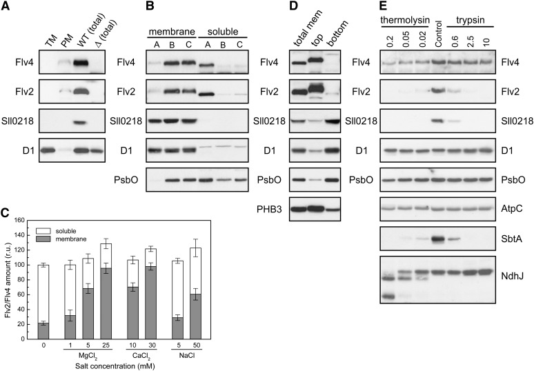
Cellular Locations of the Flv4 (Sll0217), Sll0218, and Flv2 (Sll0219) Proteins in Synechocystis.
(A) Thylakoid membrane (TM) and plasma membrane (PM) were purified from wild-type total membranes according to Norling et al. (1998), and total membranes from the wild type (WT [total]) and Δsll0217-18 mutant (Δ [total]) were isolated as described in Methods.
(B) Comparison of the distribution of the Flv2, Sll0218, and Flv4 proteins in the membrane and soluble fractions isolated by different buffer systems: buffer A (no Mg2+ or Ca2+), buffer B (30 mM CaCl2), and buffer C (25 mM MgCl2 was supplemented in buffer A).
(C) Relative distribution of the Flv2 and Flv4 proteins in the membrane and soluble fractions upon breaking the wild-type cells in the isolation buffer supplemented with different concentration of MgCl2, CaCl2, and NaCl. The amount of proteins was shown in relative units (r.u.), and the amount obtained by the buffer with no cation was set as 100. Flv2 and Flv4 proteins showed similar partitioning, which is highly dependent on the concentration of Mg2+ and Ca2+ ions.
(D) Distribution of the Flv4, Sll0218, and Flv2 proteins in the membrane fraction. Total membranes (mem) were isolated using buffer D (25 mM MgCl2) and further fractionated by a modified two-phase partitioning system as described in Methods.
(E) Stability of the Flv4, Sll0218, and Flv2 proteins in the membrane subjected to proteolysis by different concentration of trypsin (µg/mL) or thermolysin (mg/mL).
The cells were grown at air level of CO2. The Flv4, Sll0218, and Flv2 proteins were detected by protein immunoblot. D1 and PsbO of PSII were marker proteins of the thylakoid membrane and the lumen, respectively, and prohibitin 3 (PHB3) was used as a marker protein of the plasma membrane.

