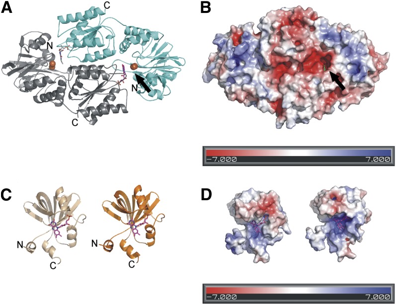
Figure 8.
Homology Model of the Flv2/Flv4 Heterodimer.
(A) The overall fold of the Flv2/Flv4 heterodimer. The functional reactive site is seen on the right with FMN (magenta) from the Flv2 monomer (gray) and diiron site from the Flv4 monomer (cyan). The irons are shown as orange spheres.
(B) The electrostatic surface of the Flv2/Flv4 heterodimer. The FMN (green) is visible in the reactive site cavity.
(C) The homology models of the flavin reductase domains with bound FMN; Flv2 is to the left (beige) and Flv4 to the right (orange).
(D) The electrostatic surface of the flavin reductase domains.

