Figure 7.
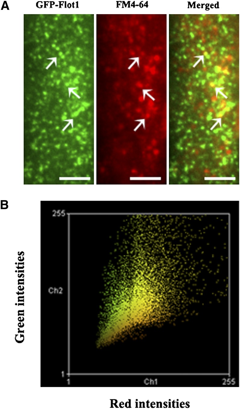
GFP-Flot1 and FM4-64 Puncta Are Often Colocalized at the Plasma Membrane of the Root Epidermal Cells.
(A) VA-TIRFM image of a root epidermal cell expressing GFP-Flot1 treated with FM4-64. White arrows indicate examples of colocalized puncta.
(B) Colocalization histogram of a plot for each pixel according to its red (x axis) and green (y axis) intensity. These data were analyzed using Manders coefficients.
Bars = 5 μm.

