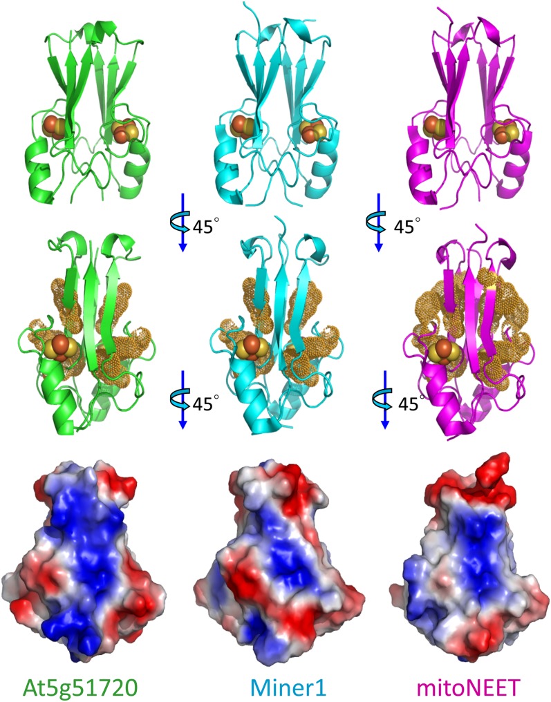
Figure 2.
Structural Comparison of At-NEET (At5g51720) with the Human Miner1 and mitoNEET 2Fe-2S Cluster–Containing NEET Proteins.
The top row shows the secondary structure of the different proteins with the 2Fe-2S centers shown as spheres (Fe is red and S is yellow). The middle row highlights the aromatic groups. Note that this view is rotated 45° from the top view to better illustrate the aromatic side chain distribution. The bottom row shows the overall surface charge based on vacuum electrostatics. All proteins are net positively charged at neutral pH and all are positively charged in the middle. By contrast, they all differ in the extent of continuous positive charge. Note that this view is rotated an additional 45° from the middle view to better illustrate the differences in the surface charges. At5g51720, green; human Miner1, cyan; mitoNEET, magenta.

